Confocal superresolution FLIM microscopy

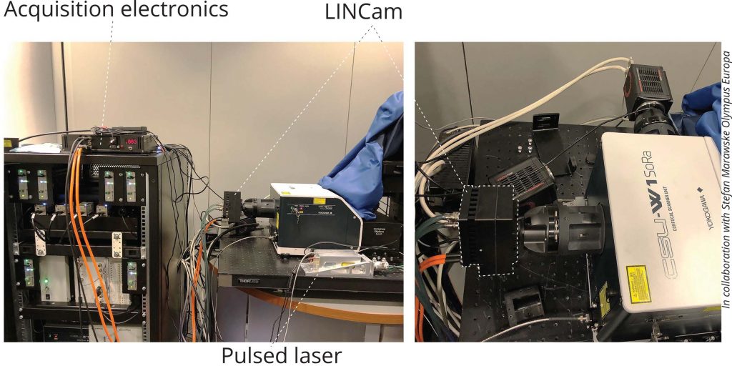
Photonscore LINCam used here as a drop-in replacement of the Hamamatsu CCD camera. The detector housing has a c-mount female thread that allows a direct attachment to the CSU. Acquisition electronic module controls the camera and transfers acquired data to the computer with a standard USB interface. A pulsed laser is required to perform time-resolved single photon counting. Here, Omicron QuixX 488 diode laser was coupled into a light input of CSU using a single mode fibre.
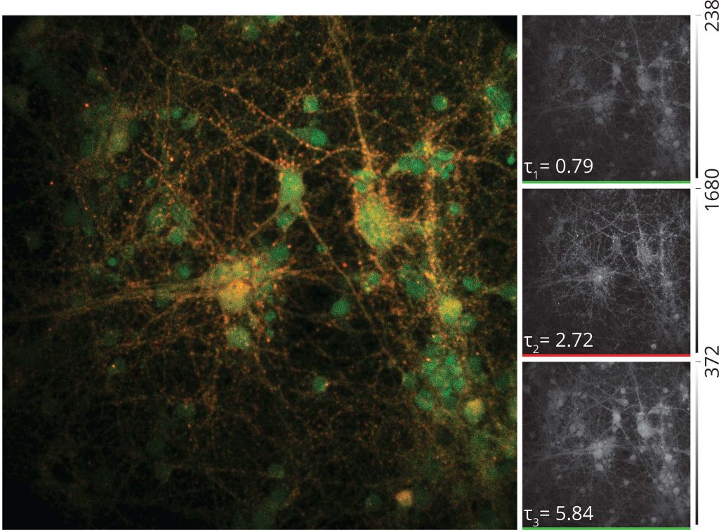
Primary hippocampal neurons from rats. To visualize excitatory synaptic contacts, neurons were stained with rat anti-homer, guinea pig anti-MAP2, rat anti-Ctip2 and mouse anti- Prox1 antibodies. Subsequently, samples were incubated with anti-rat Alexa 488-, anti-guinea pig Cy5-, and anti-mouse Alexa 350- conjugated donkey secondary antibodies.


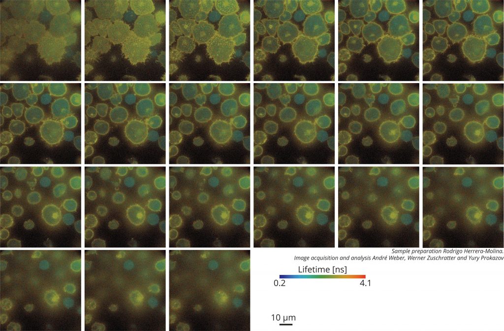
Lymphocytes (Jurakt T-cells) were transfected with a monomeric CFP-YFP Lck-biosensor and stimulated by CD3. After fixation cells were immune stained by an anti-GFP antibody, labelled with Atto 647N. The optical sections clearly show the shuttling of Lck positive vesicle between the plasma membrane and an inner compartment (most likely associated with the Golgi complex). Intensity weighted 3D stack of FLIM images of T-cells, 400 × 400 bins, 20 seconds per slice.
