One of the well-known drawbacks of widefield microscopy imaging its low axial resolution compared to confocal microscopy. Several methods are known to overcome those limitations introducing confocality. Here we demonstrate FLIM images acquired with optical sectioning with light-sheet illumination.
Image acquisition and analysis by Ovliver Kobler, André Weber and Werner Zuschratter.
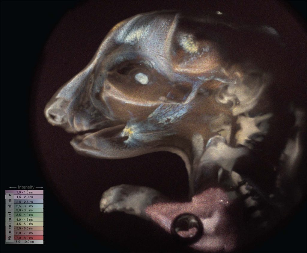
The recorded slices of the specimen can be combined into a real 3D representation of the FLIM data, allowing it to get a vivid visualization of the sample
Drosophila larvae
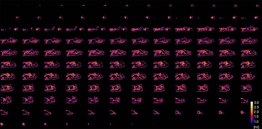
A commercially available light-sheet system equipped with a pulsed laser source and a CCD with c-mount. For the user it is just a drop-in replacement of the CCD by LINCam to start imaging. An image below shows 128 FLIM sections acquired with 10 seconds per frame.
Again the recorded data can be fused into a 3D representation.