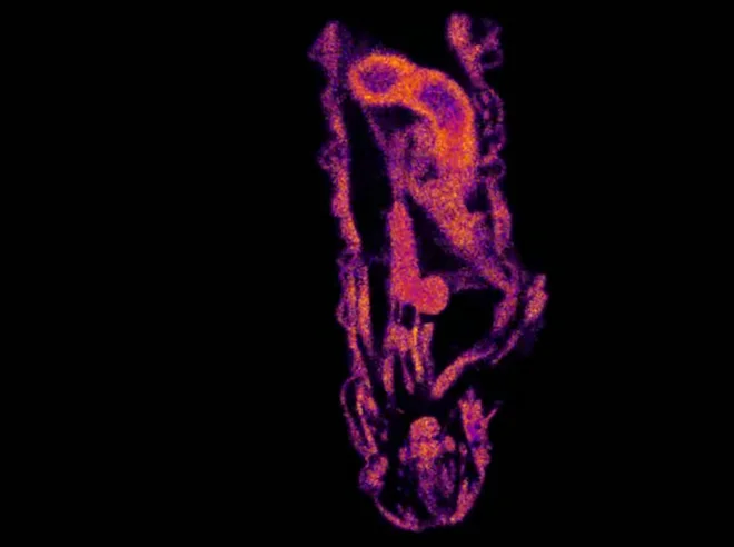
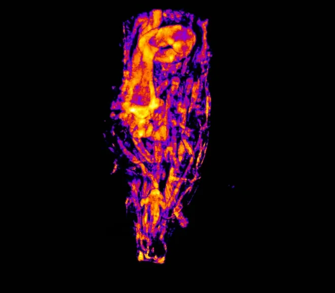
LINCam makes confocal light-sheet microscopy remarkably easy to adopt. Because it drops straight into a standard C-mount camera port, upgrading an existing light-sheet system to fluorescence-lifetime imaging is as simple as swapping the CCD for LINCam — no realignment or rebuild required. Paired with light-sheet illumination, LINCam delivers optically sectioned FLIM with the high axial resolution of confocal imaging, letting users record hundreds of lifetime-resolved slices and combine them into vivid 3D reconstructions. The result is fast, gentle, high-contrast lifetime imaging that makes confocal light-sheet FLIM genuinely pleasant to use.
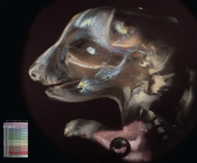
Rat embryo acquired with light-sheet microscope
One of the well-known drawbacks of widefield microscopy imaging is its low axial resolution compared to confocal microscopy. Several methods are known to overcome those limitations by introducing confocallity. Here we demonstrate FLIM images acquired with optical sectioning with light-sheet illumination.
The recorded slices of the specimen can be combined into a real 3D representation of the FLIM data, allowing it to get a vivid visualization of the sample.
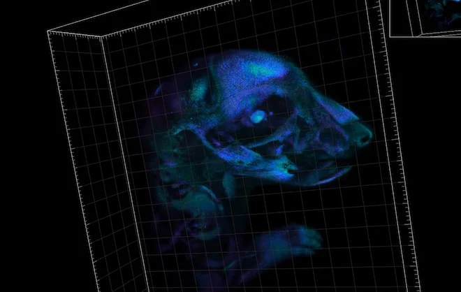
Drosophila larvae
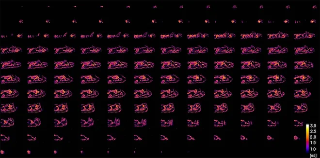
A commercially available light-sheet system equipped with a pulsed laser source and a CCD with c-mount. For the user it is just a drop-in replacement of the CCD by LINCam to start imaging. An image below shows 128 FLIM sections acquired with 10 seconds per frame.